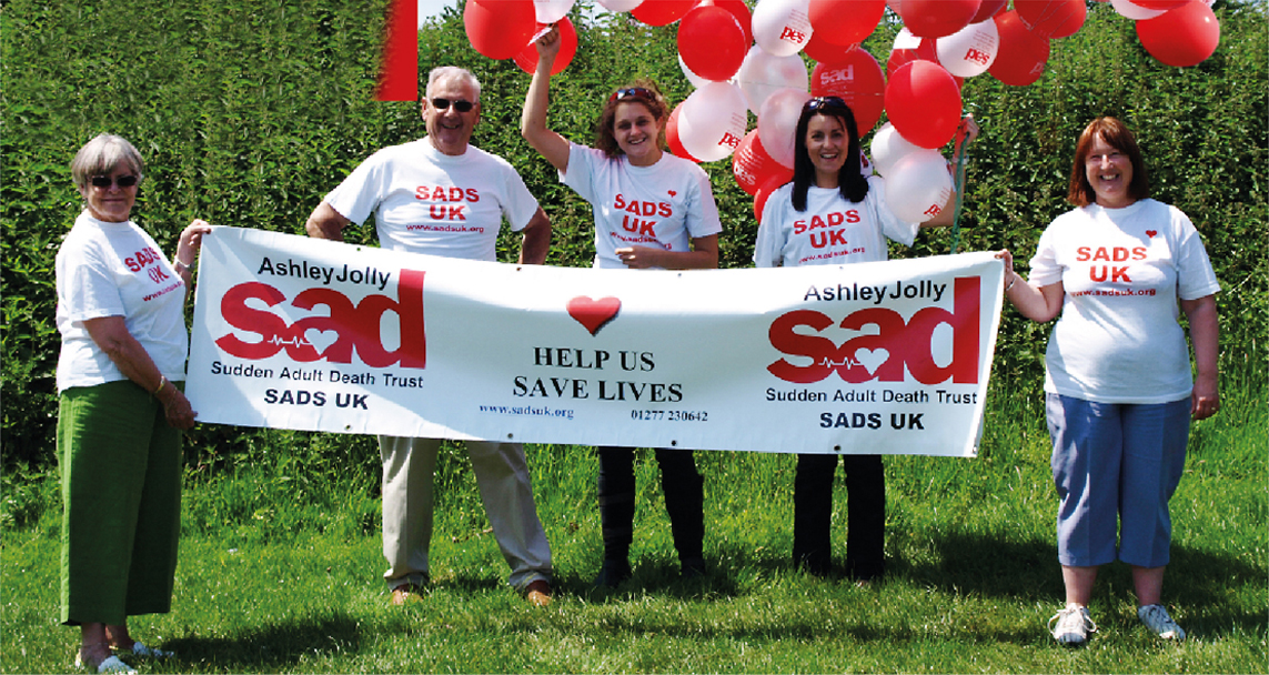
Contact SADS UK for details of our SADS memorial candle lighting, Annual Retreat and Stride to STOP SADS
Contact SADS UK for details of our SADS memorial candle lighting, Annual Retreat and Stride to STOP SADS
The heart is divided in to four chambers, two upper chambers called the atria and two lower chambers called the ventricles. The right side of the heart supplies the lungs via the pulmonary artery and the left side supplies the rest of the organs via the aorta. Situated in the right atrium are the sinoatrial (SA node) cells, these cells are source of the electrical signal controlling the heartbeat.
The heartbeat starts when the electrical signal from the SA node causes the two upper chamber muscles of the heart to contract. The blood in the atria is then forced through the atrio-ventricular AV valves in to the two lower chambers. The atrio-ventricular valves (mitral valve, tricuspid valve) close to only allow fluid to pass in one direction, this prevents blood returning to the atria as the ventricles contract. The ventricles contract when the electrical signal that started in the SA node passes through the AV node to the ventricle chamber muscles. The blood is then forced through the aortic and pulmonic valves into the arteries. The aortic and pulmonic valves close again only allowing fluid to pass in one direction, they are situated one on the left side of the heart and the other on the right side. The when these valves close, the heart muscles relax allowing the chambers to expand and the atria to refill with blood from the veins ready for the start of the next beat.
Just enter your details below

Thank you for considering donation to SADS UK.
There are several methods by which we can accept donations.
FIND OUT MORE HERE